
MRI
VEI has been in partnership with the Marion duPont Equine Medical Center since April 2004 to offer magnetic resonance imaging (MRI) directly to our clients. Since that time, hundreds of horses have been examined predominantly for foot problems. MRI provides images with unmatched tissue contrast and anatomic definition, offering significant diagnostic advantages when ultrasonography and radiography are unable to provide a clear diagnosis.
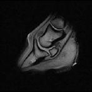
MRI displays anatomic and physiologic detail in both bony and soft tissue structures through a series of tomographic slices using magnetic properties of the horse’s tissues. MRI has provided the diagnosis in many cases when other imaging modalities failed to clearly identify the source of the lameness within the hoof capsule. This is especially true for soft tissue injuries around joints, and in the areas difficult to palpate or image by other means such as radiographs or ultrasound. MRI is the only method presently available that can assess all tissues during a single examination.
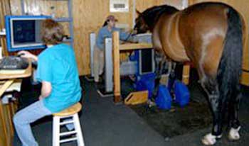
The equipment at the Marion duPont Equine Medical Center is a standing open MRI system, which allows the patients to stand under light sedation, so distal extremities can be scanned in a weight-bearing state. This enables our doctors to more precisely pinpoint your horse’s source of pain. In order to obtain images, a receiving coil is placed closely along the horse’s anatomic region of interest to collect emitted signal. The patient’s legs are positioned within the center of a strong magnetic field generated by the MRI system. The sequences are selected, and a radio frequency signal is collected to create the image. The typical MRI exam of an equine patient yields 300-500 high-detailed images to review.
MRI should be considered as a diagnostic tool when the site of pain or injury can be localized, but the problem cannot be distinguished by other forms of imaging such as radiographs or ultrasound. For instance, if your horse has been diagnosed with navicular disease, but has been unresponsive to therapy, the MRI may be the next logical approach to further characterize the structures involved and extent of injury.
The MRI exam, because it can more precisely evaluate soft tissue and bone within the hoof capsule, provides a more concise diagnosis such as navicular bursitis, impar ligament desmitis, or supensory ligament desmitis of the navicular bone. It is particularly useful for lameness localized to the foot. Importantly, MRI is capable of demonstrating cortical erosions along the flexor surface of the navicular bone, and adhesions to the adjacent deep digital flexor tendon. The use of MRI in equine veterinary medicine allows more timely intervention and an improved prognosis and long-term outcome for equine athletes.
MRI has become one of the most important diagnostic tools in equine lameness. For further information concerning specifics of the procedure or in order to make an appointment please consult our website.