Balance Films
A balanced foot is the foundation for a sound horse.
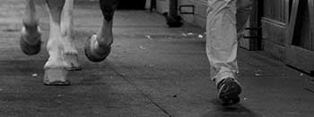
Unchecked Hoof Problems
There are many common, subtle problems of the hoof and foot, that if left unchecked, can progress over time to cause lameness in an otherwise healthy horse. Under-run or contracted heels, long toes, broken back hoof-pastern angles, flared hoof wall, toed in or out feet, and negative palmar angles of the coffin bone are some of these problems. At Virginia Equine Imaging we understand that an important part of preventing, diagnosing, and treating these problems pertaining to the feet is to start by looking at the bones within the hoof capsule through radiography.
Service Collaboration
The veterinarians at Virginia Equine Imaging frequently work closely with farriers to obtain the best results for your horse. To facilitate collaboration, we can easily send your horse’s balance films electronically for your farrier to review. With a solid foundation on all four feet, your horse has the opportunity to reach its full potential. We recommend these radiographs be taken every 6 months to maintain the best shoeing possible.
Assessing Balanced Hooves
We use state-of-the-art digital radiography to obtain lateromedial and dorsopalmar views, also called “balance films”, of the foot. Balance films allow us to visualize the bony column in relation to the external structures including the hoof wall, sole, and heels. With this additional information we can more accurately assess that your horse is distributing weight evenly over the hoof and not putting unnecessary strain on the bones and soft tissue structures of the distal limb, including the coffin joint, navicular bone and deep digital flexor tendon.
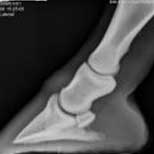
Radiograph 1: A horse with a normal hoof-pastern angle. The angles of the coffin bone and the short pastern bone are the same.
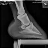
Radiograph 2: A horse with a broken back hoof-pastern angle. The angle of the short pastern bone is more upright than the angle of the coffin bone. Although this horse does not have a negative palmar angle (this is when the coffin bone is closer to the ground at the heel rather than at the toe) this
Read More..
often occurs along with a long toe, low heel, and broken back angle conformation. The result is increased pressure and concussion on the navicular bone and navicular bursa, increased tension on the deep digital flexor tendon, and more force required for breakover. These horses can become chronically heel sore.
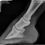
Radiograph 3: A horse with a broken forward hoof-pastern angle. The angle of the coffin bone is more upright than the angle of the short pastern bone. This conformation puts more stress on the coffin joint and can lead to coffin joint effusion and synovitis.
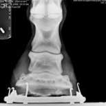
Radiograph 4: A horse with medial-lateral imbalance. Notice that the lateral (outside) hoof wall is higher than the medial (inside) hoof wall. This can cause the heels, distal interphalangeal (coffin) joint, proximal interphalangeal (pastern) joint, and metacarpophalangeal (fetlock) joint to suffer uneven compressive forces.
