Virginia Equine Imaging

Meet Our Team
We are pleased to offer comprehensive services including decades of combined experience in lameness and performance concerns ranging from pleasure horses to FEI competitors, as well as expertise in neurology, gastrointestinal, and airway medicine to evaluate and manage the whole horse. We offer both in clinic appointments and farm calls.
Diagnostic Imaging
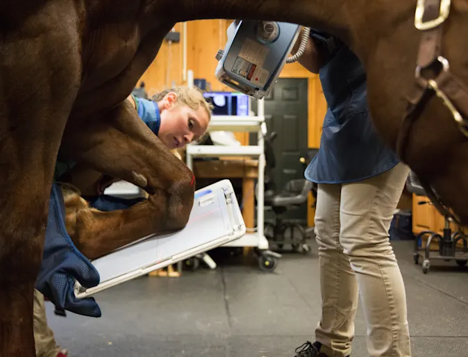
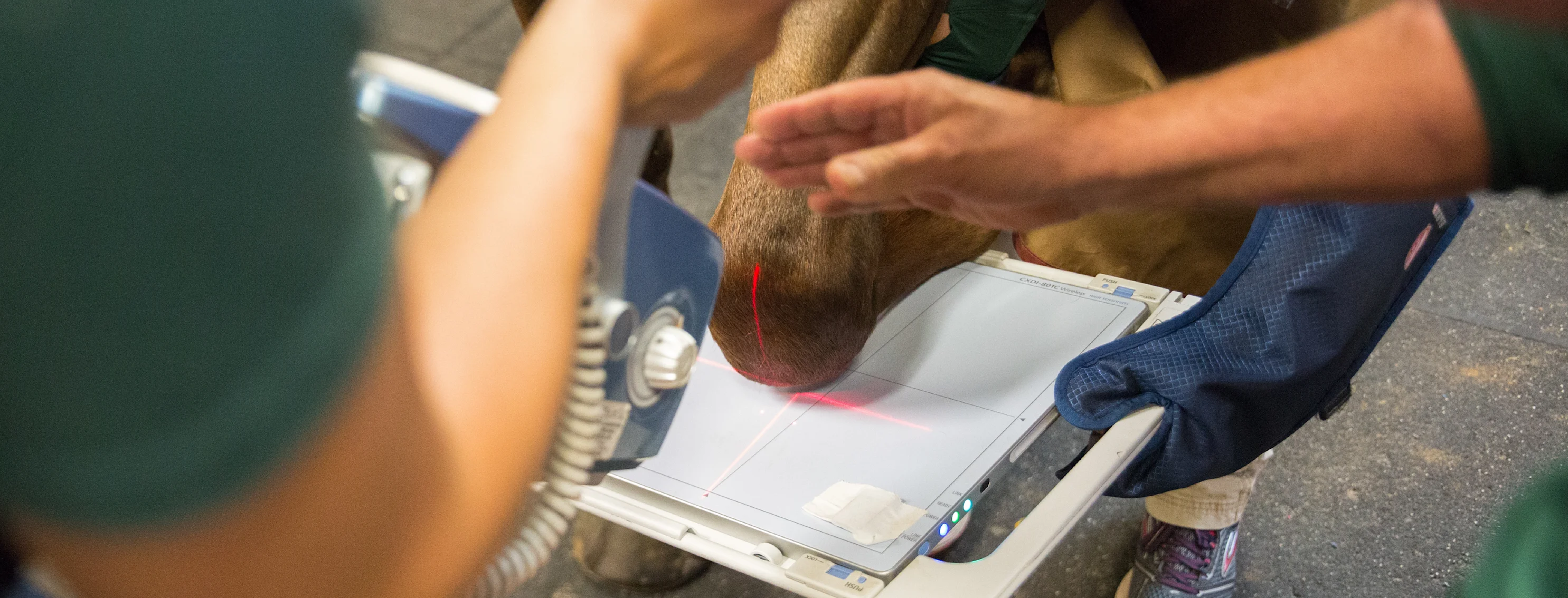
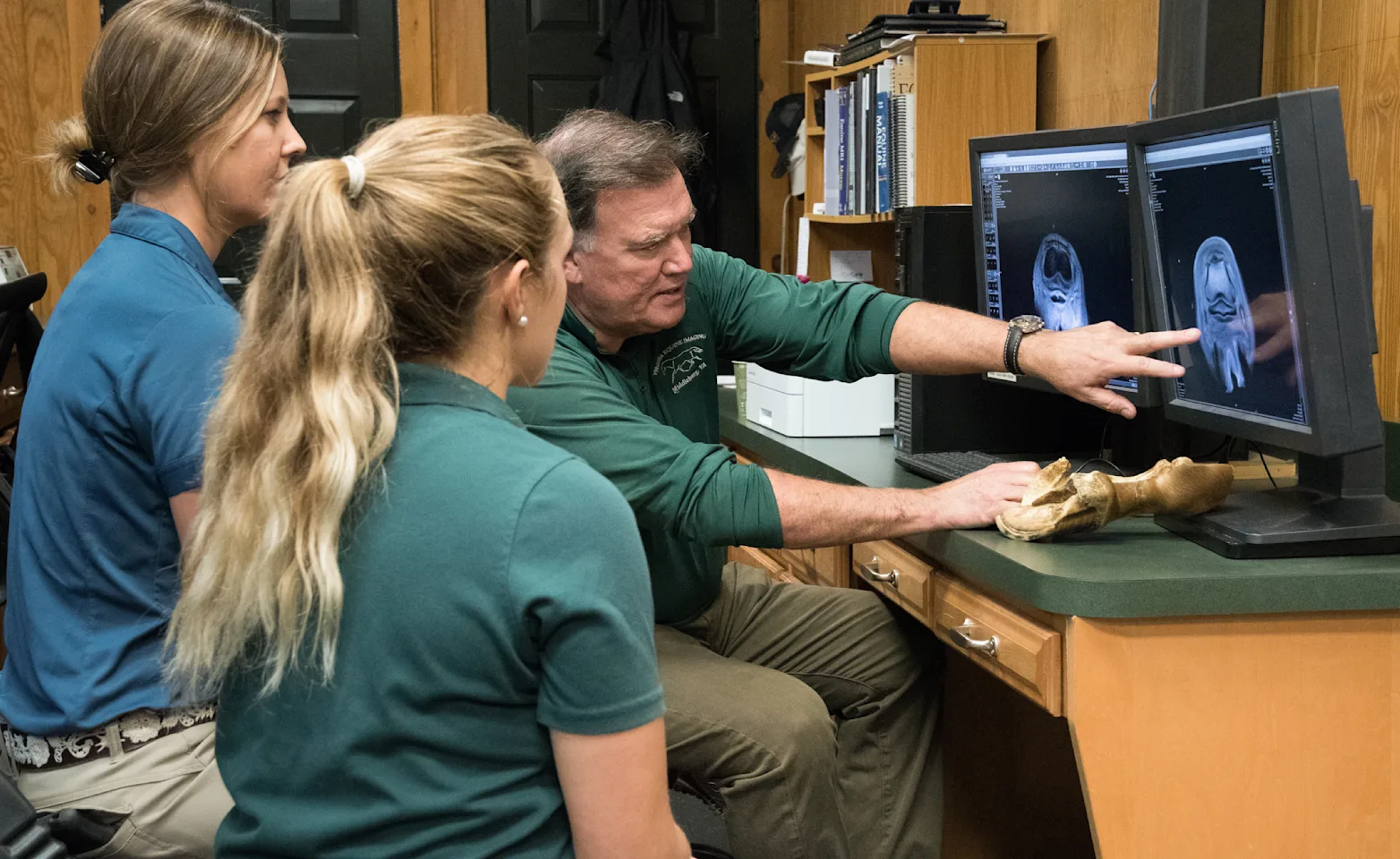
Diagnostic imaging allows veterinarians to see inside a horse’s body without the need for surgery. X-rays are probably the best-known type of diagnostic imaging, but many others are available to help diagnose illnesses and other health problems in horses.
Each type of diagnostic imaging has its own advantages and disadvantages. None of these are perfect for every situation. Your veterinarian will suggest one that best fits the needs of your horse. Some of these tests are available in most veterinary practices, but others may require a visit to a special veterinary diagnostic-imaging clinic.
Client Reviews & Testimonials
We value our clients’ experience at Virginia Equine Imaging. Here’s what some people are saying about us.
Wonderful facility, an excellent stack is excellent staff all the way around. They made me and my horse feel welcome and I feel very good about the diagnosis and treatment that they offered my horse.
Ann B.
Unparalleled service and expertise. Any horse would be lucky to have this caliber of care.
C.L.
Always have a great experience with the team at VEI. Everyone is nice and puts the horse first. The facilities are clean and comfortable for both humans and horses. Dr. Allen and the team got to the bottom of my horse's issues and set a road map to success. Happy to say all is well and now will hopefully only see them for a yearly exam.
Michele
